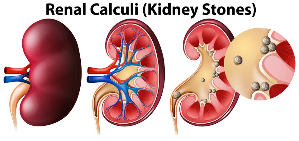
Kidney stones, medically known as renal calculi or nephrolithiasis, are hard deposits of minerals and salts that form inside the kidneys. They are among the most common urological disorders worldwide and affect approximately 10-15% of people during their lifetime. Kidney stones can cause severe pain, urinary symptoms, and complications if left untreated. While many stones pass naturally, larger stones may require medical intervention. Scientific evidence suggests that dietary modifications, adequate hydration, and appropriate medical treatment can significantly reduce the risk of stone formation and recurrence.
What Are Kidney Stones?
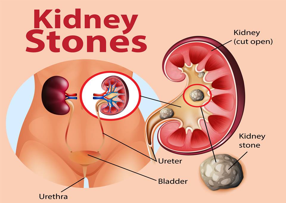
Kidney stones are solid crystalline masses formed when certain substances in urine, such as calcium, oxalate, uric acid, and cystine, become highly concentrated and crystallize. These crystals may grow over time and develop into stones of varying sizes.
The most common types include:
- Calcium oxalate stones (70-80% of cases)
- Calcium phosphate stones
- Uric acid stones
- Struvite stones (associated with urinary tract infections)
- Cystine stones (caused by genetic disorders)
Research published in the New England Journal of Medicine has shown that stone recurrence is common, with nearly 50% of patients experiencing another stone episode within 5–10 years if preventive measures are not implemented (Curhan et al., 1993).
Symptoms of Kidney Stones
The symptoms of kidney stones depend on their size, location, and whether they obstruct urine flow. Small stones may pass unnoticed, while larger stones can cause intense discomfort.
Common symptoms include:
- Severe pain in the back, side, lower abdomen, or groin
- Pain that comes in waves and fluctuates in intensity
- Burning sensation during urination
- Blood in urine (hematuria)
- Cloudy or foul-smelling urine
- Frequent urge to urinate
- Nausea and vomiting
- Fever and chills if infection is present
The classic symptom is renal colic, often described as one of the most severe forms of pain. Studies published in The Lancet have reported that ureteral obstruction caused by stones triggers intense spasms and pressure buildup, leading to characteristic renal colic pain (Türk et al., 2024).
How to Diagnose Kidney Stones
Accurate diagnosis is essential for determining stone size, location, composition, and the most appropriate treatment strategy.
A physician may recommend:
Medical History and Physical Examination
Healthcare providers assess symptoms, family history, dietary habits, hydration status, and previous stone episodes.
Urine Testing
Urinalysis helps detect:
- Blood in urine
- Infection
- Crystal formation
- Urinary pH abnormalities
Twenty-four-hour urine testing is often recommended for recurrent stone formers to identify metabolic risk factors.
Blood Tests
Blood investigations may measure:
- Calcium
- Uric acid
- Creatinine
- Electrolytes
These tests help identify underlying conditions contributing to stone formation.
Imaging Studies
Non-contrast computed tomography (CT) is considered the gold standard for kidney stone diagnosis due to its high sensitivity and specificity. Ultrasonography is commonly used in children and pregnant women to avoid radiation exposure.
The American Urological Association (AUA) guidelines recommend CT imaging as the preferred diagnostic tool for most adults with suspected kidney stones (Assimos et al., 2016).
How to Prevent Kidney Stone Formation
Preventing kidney stones is often more effective than treating recurrent episodes. Lifestyle and dietary modifications have been shown to substantially reduce recurrence risk.
Stay Well Hydrated
Adequate fluid intake is the most effective preventive strategy. Increased urine volume dilutes stone-forming substances and reduces crystal formation.
A landmark randomized clinical trial published in the New England Journal of Medicine found that patients maintaining urine output above 2 liters daily experienced significantly fewer recurrent stones compared with controls (Borghi et al., 1996).
Most adults should aim for:
- 2.5-3 liters of fluid daily
- Urine that remains pale yellow in color
Maintain Adequate Dietary Calcium
Contrary to popular belief, restricting dietary calcium may increase stone risk. Calcium binds oxalate in the intestine and reduces oxalate absorption.
A randomized trial demonstrated that a normal-calcium diet combined with reduced sodium and animal protein intake lowered recurrence rates more effectively than a low-calcium diet (Borghi et al., 2002).
Limit Excess Sodium
High sodium intake increases urinary calcium excretion, promoting calcium stone formation. Clinical guidelines recommend limiting sodium intake to less than 2,300 mg per day.
Moderate Animal Protein Consumption
Excessive consumption of red meat and animal proteins can increase urinary calcium, uric acid, and oxalate levels while reducing protective citrate levels.
Reduce High-Oxalate Foods in Susceptible Individuals
Individuals prone to calcium oxalate stones may benefit from moderating intake of:
- Spinach
- Beetroot
- Rhubarb
- Nuts
- Chocolate
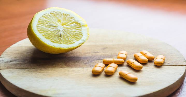
Increase Citrate-Rich Foods
Citrate inhibits crystal formation and stone growth. Citrus fruits such as lemons and oranges are natural sources of citrate.
Research published in Urology reported that citrate supplementation may help reduce stone recurrence in selected patients (Barcelo et al., 1993).
How to Remove Kidney Stones
Treatment depends on stone size, location, symptoms, and complications.
Conservative Management
Many stones smaller than 5 mm pass naturally with:
- Increased fluid intake
- Pain-relieving medications
- Medical observation
Studies indicate that approximately 68-98% of stones smaller than 5 mm pass spontaneously (Miller & Kane, 1999).
Medical Expulsive Therapy
Alpha-blockers such as tamsulosin may relax ureteral muscles and facilitate stone passage.
A meta-analysis published in European Urology found that alpha-blockers improved spontaneous stone passage rates, particularly for larger ureteral stones (Hollingsworth et al., 2016).
Extracorporeal Shock Wave Lithotripsy (ESWL)
ESWL uses focused sound waves to break stones into smaller fragments that can pass through urine.
It is commonly used for:
- Stones less than 2 cm
- Stones located in the kidney or upper ureter
Ureteroscopy
A thin scope is inserted through the urinary tract to visualize and remove or fragment stones using laser technology. This procedure is highly effective for ureteral stones and larger calculi.
Percutaneous Nephrolithotomy (PCNL)
PCNL is recommended for:
- Large stones (>2 cm)
- Complex or multiple stones
- Staghorn calculi
Clinical studies demonstrate stone-free rates exceeding 85–90% following PCNL in appropriately selected patients (Türk et al., 2024).
When to Seek a Doctor
Immediate medical attention is necessary if any of the following symptoms occur:
- Severe or persistent pain
- Fever or chills
- Difficulty passing urine
- Persistent nausea and vomiting
- Blood in urine
- Signs of urinary tract infection
- Single functioning kidney with suspected obstruction
Untreated obstruction combined with infection can become a medical emergency and may lead to kidney damage or sepsis.
Patients with recurrent stones should also seek medical evaluation for metabolic testing and long-term prevention strategies.
Bottom Line
Kidney stones are a common but often preventable urinary tract condition. They develop when minerals and salts crystallize within the kidneys and may cause severe pain, urinary symptoms, and complications. Modern diagnostic tools such as CT imaging allow accurate detection, while treatments ranging from hydration and medications to minimally invasive procedures can effectively remove stones. Strong clinical evidence shows that adequate hydration, maintaining normal dietary calcium, reducing sodium intake, and following individualized dietary recommendations can significantly reduce recurrence risk. Early diagnosis and preventive strategies remain the most effective approaches for long-term kidney stone management.
References
- Assimos D, Krambeck A, Miller NL, et al. Surgical Management of Stones: American Urological Association Guideline. Journal of Urology. 2016;196(4):1153-1160.
- Barcelo P, Wuhl O, Servitge E, Rousaud A, Pak CY. Randomized double-blind study of potassium citrate in idiopathic hypocitraturic calcium nephrolithiasis. Journal of Urology. 1993;150(6):1761-1764.
- Borghi L, Meschi T, Amato F, et al. Urinary volume, water and recurrences in idiopathic calcium nephrolithiasis: A randomized trial. New England Journal of Medicine. 1996;334(1):77-80.
- Borghi L, Schianchi T, Meschi T, et al. Comparison of two diets for the prevention of recurrent stones in idiopathic hypercalciuria. New England Journal of Medicine. 2002;346(2):77-84.
- Curhan GC, Willett WC, Rimm EB, Stampfer MJ. Family history and risk of kidney stones. Journal of the American Society of Nephrology. 1997;8(10):1568-1573.
- Hollingsworth JM, Canales BK, Rogers MA, et al. Alpha-blockers for treatment of ureteric stones: Systematic review and meta-analysis. European Urology. 2016;69(6):1099-1107.
- Miller OF, Kane CJ. Time to stone passage for observed ureteral calculi. Urology. 1999;53(4):688-691.
- Türk C, Petrik A, Sarica K, et al. EAU Guidelines on Urolithiasis. European Association of Urology Guidelines. 2024.
- Written By: Shabina Khan (Clinical Pharmacist)
- Medically Reviewed By: Dr. Prateek Sharma (Advisor Health & Wellness)